


Search websites, locations, and people

School of Life Sciences ACADEMICS
Researchers at Westlake University Unveiled Structure of the RBD-ACE2-B0AT1 Complex
25, 2020
Email: fengyi@westlake.edu.cn
Phone: +86-(0)571-85270350
Office of Public Affairs
Science published the research paper titled Structural basis for the recognition of the SARS-CoV-2 by full-length human ACE2 online via First Release around 10am EST on March 4th. In this paper, researchers from Qiang Zhou's Lab of westlake University report the full-length structure of ACE2, the receptor of the novel coronavirus SARS-CoV-2, as well as the 3D structure of the full-length human ACE2-B0AT1 complex in apos and the RBD of 2019-nCoV in its bounded state.
(Check the following website to read the full article.
https://science.sciencemag.org/content/early/2020/03/03/science.abb2762)
These two papers were previously published on bioRxiv on Feb 19th and Feb 21st respectively so that the information could be accessed by the public.
What do we know about ACE2 and the S-protein?
The outbreak of COVID-19 added new biological terminology such as coronavirus, S-protein and ACE2 into our daily-life vocabulary. ACE2 has been suggested to be the cellular receptor for the novel coronavirus. Like other coronaviruses, such as the SARS-CoV, the 2019-nCoV uses the RBD of its surface’s spike glycoprotein (S-protein) to bind with ACE2. The S-protein, which can be considered an enemy of the human body, is trying to hijack ACE2 and invade the human body through binding with it..
The S-protein, is located on the surface of the 2019-nCoV, forming its crown-like appearance. According to the latest research published by researchers at UT Austin, the S-protein of the 2019-nCoV is assembled as a trimer, containing around 1,300 amino acids within each unit. The receptor binding domain (RBD) of the S-protein, which contains around 300 amino acids, mediates the binding with ACE2. ACE2, an innocent protein whose daily responsibility is to lower the blood pressure, exists widely in the human lung, heart, kidney and intestine.
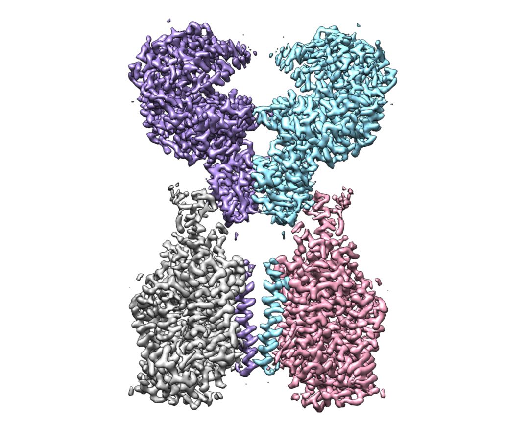
Cryo-EM map of the ACE2-B0AT1 complex.
How does it look like, when a human body protein interacts with a virus? Liang Tao, a Principle Investigator at Westlake University offered an analogy: "If we think of the human body as a house and 2019-nCoV as a robber, then ACE2 would be the doorknob of the house’s door. Once the S-protein grabs it, the virus can enter the house."
How does the S-protein bind with ACE2?
Though the S-protein and ACE2 are at the frontline of the contact, scientists have never fully understood ACE2 or its interaction with S-proteins.
Two days ago, Qiang Zhou’s lab released the first high-resolution 3D-structure of the full-length human ACE2. They will also unveil the structure of the full-length human ACE2 bound to the RBD of the 2019-nCoV soon. The overall resolution of the complex is 2.9Å, while the local resolution at the ACE2-RBD interface is 3.5Å.
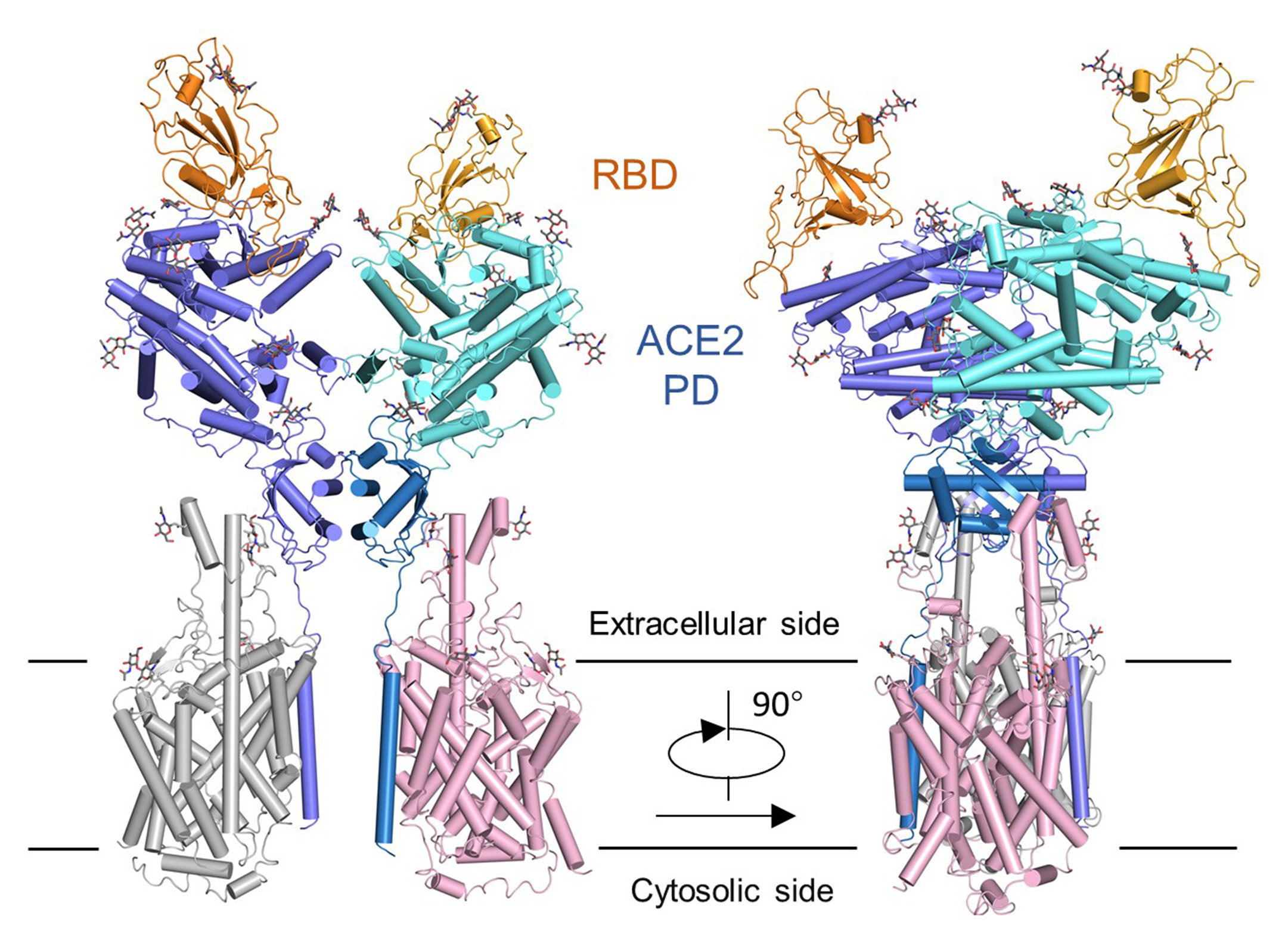
Overall structure of the RBD-ACE2-B0AT1 complex.
So, what did they see from the ternary structure?
They found that the RBD of S-protein is spanned above the surface of ACE2 like a bridge. It also looks like an evil hand of the virus getting a strong hold of the ACE2. The overall interface is similar to that between the SARS-CoV and ACE2, whereas more variations have been observed. The RBD of the S-protein on the 2019-nCoV shares about 82% similarity with the ones of the SARS-CoV.
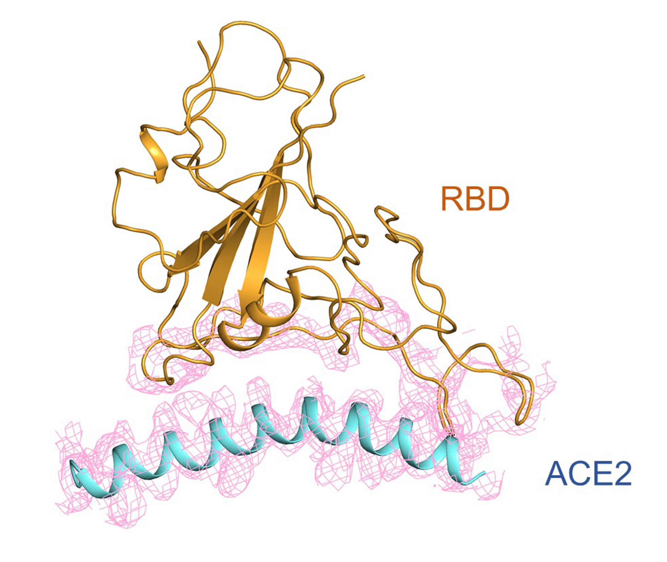
Cryo-EM density of the interface between RBD of the S protein of 2019-nCoV and ACE2.
Through further analysis, researchers figured out what amino acids of the S-protein are involved in the interaction with ACE2. Comparing to the known interactive mechanism between SARS-CoV and ACE2, some of the leftover amino acids imply obvious differences of the S-protein of the 2019-nCoV. This could potentially explain why it has a different binding with ACE2 than that of SARS. This unique binding could impact the infectious capability of the virus, but the difference of binding affinity requires further methods to measure.

Structure comparison between the RBD-ACE2-B0AT1 ternary complex and the spike glycoprotein. The structure of the S protein is superimposed to the RBD of the RBD-ACE2-B0AT1 ternary complex. Left: S protein of 2019-nCoV (PDB code: 6VSB). Right: S protein of SARS-CoV (PDB code: 6ACJ).
It is reported that teams led by Dr. Xinquan Wang at Tsinghua University and Dr. Jianxun Qi at the Institute of Microbiology, Chinese Academy of Sciences, have respectively analyzed the crystal structure of the complex between the n-terminus protease domain of ACE2 and the RBD of the S-protein of 2019-nCoV. Their research complements with the Cryo-EM structure discovered by Dr. Zhou’s team. It shall be noticed that all three teams chose to share the atomic coordinates of the complex with all people so as to speed up the therapeutic methods against 2019-nCoV.
To download the atomic coordinates of the work of Dr. Qiang Zhou’s team at Westlake University on the complex of the full-length ACE2 bounding to the RBD of 2019-nCoV, please visit: https://www.jianguoyun.com/p/DVLSdMkQiJ2OCBjbwt8C
To download the atomic coordinates of the work of Dr. Xinquan Wang’s team at Tsinghua University on the complex of ACE2 protease ECD and S-protein ECD of the 2019-nCoV, please visit: https://www.jianguoyun.com/p/DTJ03HoQ3MX2BhjiwN8C
To download the atomic coordinates of the work of Dr. Jianxun Qi at the Chinese Academy of Sciences on the complex of ACE2 protease ECD and S-protein ECD of the 2019-nCoV, please visit: http://nmdc.cn/#/resource/detail?no=NMDCS0000001
To download the atomic coordinates of the full-length protein of ACE2 and the amino acid transporter, please visit the following links:
For closed conformation: http://nmdc.cn/#/resource/detail?no=NMDCS0000001
For open conformation: https://www.jianguoyun.com/p/DWdO-qMQiJ2OCBjGwt8C
How far off are we from the silver bullet to the COVID-19?
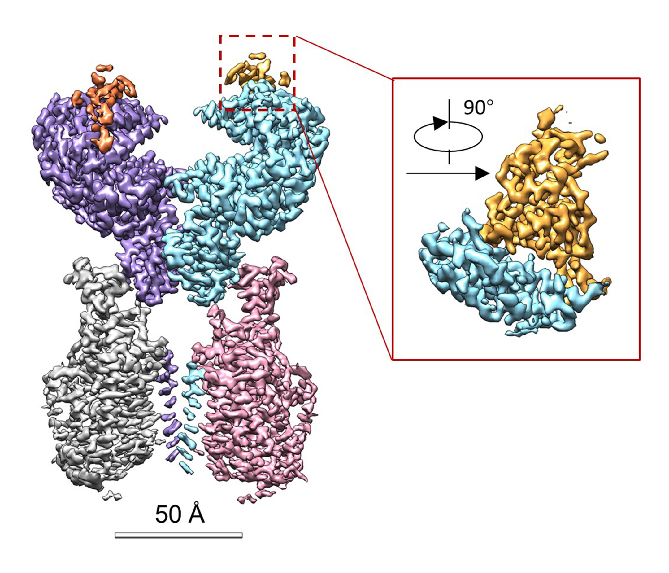
Cryo-EM map of the RBD-ACE2-B0AT1 complex. Left: Overall reconstruction of the ternary complex at 2.9 Å. Inset: focused refined map of RBD.
The COVID-19 epidemic has started to plateau, but public interest on virus-related research remains high because we are yet to find the wonder drug to COVID-19.
Dr. Zhou’s team said that their breakthrough in the complex analysis might not directly contribute to the R&D of antiviral medicine. However, it remains important because the structure of the protein determines its nature and function to a large extent. By understanding the structure of the S-protein of 2019-nCoV and ACE2, and how these two interact, we can gain a clear picture of what we are dealing with, providing more information for the scientists who are working on target medicines. It is only through sufficient information that we can achieve victory.
Another significant aspect of this research is that it offers a way for researchers in computational biology to build different models based on the structure and deploy research accordingly. It opens the door for them to decide what kind of mutation could further enhance the interaction between the S-protein and ACE2, and in turn come up with medicines and vaccinations against the interaction of S-proteins and ACE2. It could also lead to designing of small molecules to sabotage the interaction between the two. For all the options above, knowing the structure of the complex may serve as a crucial foundation for medicine and treatment developments.
For the full content of their research, please see:
Structural basis for the recognition of the 2019-nCoV by human ACE2
Renhong Yan, Yuanyuan Zhang, Yingying Guo, Lu Xia, Qiang Zhou
https://www.biorxiv.org/content/10.1101/2020.02.19.956946v1
RELATED
ACADEMICS
Researchers at Westlake University Unveiled Structure of the RBD-ACE2-B0AT1 Complex


-
STUDY WITH US
-
WORK WITH US
-
CONTACT US
-
YUNGU CAMPUS
-
No. 600 Dunyu Road, Sandun Town, Xihu District, Hangzhou, Zhejiang PR China Post code:310030 E-mail:office@westlake.edu.cn
-
-
YUNQI CAMPUS
-
No.18 Shilongshan Road Cloud Town, Xihu District, Hangzhou, Zhejiang PR China Post code:310024 E-mail:office@westlake.edu.cn
-
-
-
浙ICP备18025489号 浙公安备33010602007514号 Copyright © Westlake University. All Rights Reserved
-


If you are interested in supporting us or making a contribution, please contact:
Office of Public Affairs, Westlake University
Email: media@westlake.edu.cn
OR
Westlake Education Foundation
Email: office@wefoundation.org.cn
Phones: 0571-86886859
Fax: 0571-85271986